
Arthritis: Diagnosis
Top Diagnosis
1. Imaging tests and lab tests for osteoarthritis
X-rays show the narrowing of the space between the bones of the joint and this means loss of cartilage. Bone spurs around the bone are also seen in X-rays. Magnetic Resonance Imaging (MRI) uses radio waves and a strong magnetic field to determine the exact reason for the pain. Blood tests done in labs can also help find out the reason for the joint pain. Joint fluid analysis is done by the doctor drawing out fluid from the affected joint with a needle and the testing of the fluid will determine if the pain is due to some infection or gout.
2. Rheumatoid Factor test
Rheumatoid Factor test is done to diagnose rheumatoid arthritis. This test is ordered when the patient experiences morning stiffness in the joint, warmth, pain and swelling, and nodules under the skin. ESR or Erythrocyte Sedimentation Rate test is done to determine the existence of inflammation and the advancement of the disease in the body. CRP or C-reactive protein test id also determines the existence and advancement of the disease in the body. The higher the C-protein, the higher will be the rate of inflammation. FBC or Full Blood Count test is done as people suffering from rheumatoid arthritis also suffer from anemia.
3. Joint fluid analysis, blood test and imaging test for infectious or septic arthritis
The doctor draws out a sample of the synovial fluid from the joint and the test done on it determines what bacterium is causing the pain. The synovial fluid, which usually appears clear and thick, has an altered color if it contains bacterium. The presence of bacterium can also affect the volume, consistency and make up of the synovial fluid. A blood test is conducted to find the presence of bacterium in the bloodstream. X-rays may also be ordered to get a clear picture of the affected joint.
4. Blood tests and imaging tests for juvenile rheumatoid arthritis
Erythrocyte Sedimentation Rate (ESR) is the rate at which the red blood cells at the bottom of the blood containing tube sediment in a period of one hour. This classifies the type of juvenile rheumatoid arthritis that has attacked the joint. C-reactive protein test is also done to measure the levels of general inflammation. Rheumatoid factor test is also done. Cyclic Citrullinated Peptide (CCP) also determines the presence of an antibody in the bloodstream that can cause juvenile rheumatoid arthritis.
5. Imaging tests and lab tests for psoriatic arthritis
X-rays help to detect the changes in the joints if there is psoriatic arthritis. Magnetic Resonance Imaging (MRI) uses strong magnetic fields and radio waves that check problems in the tendons and ligaments of the joints. Rheumatoid factor and joint fluid tests are also done to confirm the attack of psoriatic arthritis.
6. Biopsies
Synovial biopsy of the knee is quite underused and little known to the general poulation. It is not only safe but can also be repeated easily. Synovial biopsy is suggested when the cause of the inflammatory joint disease still remains unclear. If the culture and examination of the synovial fluid proves unhelpful, a biopsy can help in distinguishing conditions like tuberculosis or other sub-acute infections for which some specific treatments are indicated. Biopsies are helpful when conventional tests fail in separating diseases like Whipple’s disease, amyloidosis and saocoidosis, or malignancies like polyarthritis, which are different from the usual rheumatic diseases. Biopsies are also done as some types of arthritis have skin involvement. This is used to diagnose vasculitis, lupus and even psoriatic arthritis.
7. Medical history
This involves gathering of information about the patient’s past conditions. This is done by preparing some questions. These questions give the doctors the list of medications the patient has been currently taking, list of medication allergies, a list of previous surgeries etc.
8. Hands on physical examination
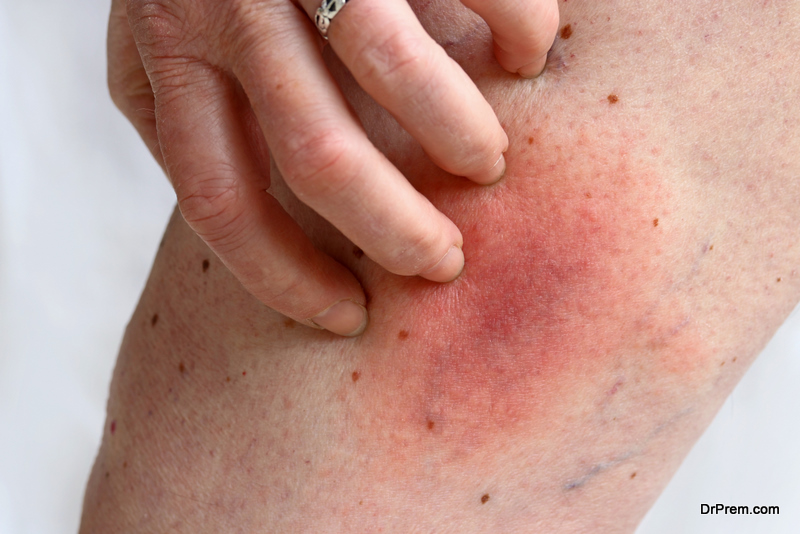
The doctor will perform a physical examination and look for visible evidence like joint stiffness, joint tenderness, warmth near the joints, bumps or nodules, fluid in a joint, redness around the joint, even fever etc to confirm the presence of arthiritis. During the examination, the doctor will take note of your range of motion to check how far it deviates from the normal range of motion for a particular joint. The doctor assesses the limitation brought about by the joint damage, muscles spasms, joint swelling etc. The examination also conveys the part you played in bringing about the damage, like the activity that had caused the joint to swell, what you had done to relieve the pain etc.
9. Blood tests
Blood tests done in the laboratory are undoubtedly important diagnostic tools. However, they are not definitive if considered alone. The HLA B27 test uses HLA B27 which is an inherited gene marker which is associated with some related rheumatic diseases. The features that these related diseases share are GI disorders, skin and peripheral arthritis, and anterior chamber eye disease. About 80% of the patients with reactive arthritis have the HLA B27 gene. However, most patients with the gene are not seen to develop any of the clinical rheumatic symptoms of significance. Blood tests are also done to check the level of uric acid in the blood as its presence results in the formation of crystals which get deposited in the joints and tissues. This gives rise to painful gout attacks. This test also determines the cause of pain in joints.