
Squamous cell carcinoma: Diagnosis
Top Diagnosis
1. Biopsy
This test is the most preferred and invasive in nature. Biopsy involves medical removal of the affected tissues and taking samples for examination. The sample is examined thoroughly by a surgeon or a radiologist to ascertain the extent of the disease. He takes a small sample of the infected tissue and treats it with dyes that stain the tissue for a more clear view. Then a pathologist examines it under a microscope to check any abnormal growth or mucus. There are different types of biopsy procedures including excisional, incisional, needle aspiration and skin biopsy. These procedures are very effective at confirming the exact nature of the cancer as well as the extent of its spread.
The biopsy specimen is examined by the pathologist under a microscope for squamous cell skin carcinoma characteristics. If any metastases are present, then the patient is said to be suffering from squamous skin carcinoma.
2. CT body scan
CT body scan or Computed Tomography is used to diagnose squamous cell carcinoma. This procedure is effective as it completely eradicates the superimposition of structures outside the area under examination. CT scan has an inherent high-contrast resolution that allows to easily distinguish the tissues with physical density less than 1 percent. CT scan is useful in detecting the extent of tumors as well as visceral nodules suggestive of metastases. This test is non-invasive and fails at evaluating the type and nature of squamous cell carcinoma.
3. MRI Scan
Magnetic Resonance Imaging (MRI) scan is a procedure that makes use of radiology to visualize the detailed internal structures of human body. It is capable of creating impressions of nuclei of atoms with utmost accuracy. This procedure involves placing the human body in a strong magnetic field. The protons within the cells align themselves in accordance to this magnetic field. As the magnetic field is switched off, these protons realign to their original positions emitting radio frequency signals. This diagnosis is very effective at evaluating the extent of the large tumors as well as to rule out metastatic disease. However, this test is not sufficient to ascertain the type and nature of cancer. Thus, it is advised to go for other tests as well.
4. PET Scan
Positron Emission Tomography is a latest technology that utilizes the gamma rays for imaging a 3D view of functional processes of the body. This is a procedure used in both medical as well as in clinical research. A positron-emitting radionuclide is used to emit the gamma rays. These rays create the 3D images with the help of a computer analysis. PET scan alongside MRI scan provides the accurate anatomic as well as metabolic functions to evaluate the spread of squamous cell carcinoma as well as to rule out metastatic disease, if any. This test is non-invasive in nature and is quite expensive.
5. FBC with differential
Full blood count (FBC) with differential is an in-depth blood examination that provides the complete information about the cells present in the blood of the patient under examination. This test is carried out by a scientist or lab technician on the prescription of a doctor. The test includes examination of white blood cells, red blood cells and blood platelets. This test is very useful at detection of distant as well as regional metastases and ascertains the risk level of the disease. During diagnosis, if any bone marrow metastases are found in the tissues under examination, then it can be a case of bone marrow cancer. This test is invasive in nature and confirms the status of the aliment.
6. LFTs
Liver Function Tests (LFTs) are about examining the state of the patient’s liver. Clinically there are certain parameters like PT/INR, albumin, Billrubin and aPTT that are checked to understand the condition of the liver. The tests also include a biopsy in which tissue samples from the kidney are retrieved using a fiber-thin tube The test is non-invasive but essential in diagnosis of both Hepatocellular carcinoma (HCC) as well as Cholangiocarcinoma, which are two types of liver cancer caused due to the squamous cell carcinoma in liver.
7. Chest X-ray
Chest X-ray is essential in case of Bronchial Catcinoma as it is helpful in detecting the presence of any metastases lesions in the lungs. Lesion is the abnormal areas of tissue on the body. If you are a chain smoker, suffer from persistent lung infection or have been losing weight drastically, then this test is recommended to ensure the presence of any metastases in the lung tissues. The neoplastic cells grow in small masses within the alveoli. The metastases lesions are present on the lymph nodes and start affecting the epithelium cells of terminal bronchioles. If there is abnormal growth of the tissues, then it can block the free flow of air in the breathing tube as well as the blood flow in the blood vessels. This test is non-invasive and requires biopsy and CT scan for understanding the complete nature of squamous cell carcinoma in lungs leading to lung cancer.
8. Fine Needle Aspiration Cytology
Fine Needle Aspiration Cytology is very effective and preferred for diagnosing primary squamous cell carcinoma in delicate internal organs of the patient’s body. In this procedure, a narrow, needle size gauge is inserted into the body to collect tissue samples of the infected area. This test allows a lesser invasive diagnosis of squamous cell carcinoma. Doctors have to carry out clinical or radiography tests to confirm the presence of squamous cell carcinoma. However, this test is useful at examining the nature of tumor along with its immunohistochemical profile. This test allows the surgeon in better prediction and prognosis. Fine needle aspiration Cytology is very sensitive at detecting both anaplastic as well as papillary carcinomas.
9. Laboratory Biochemical Testing
The presence of squamous cell carcinoma in body is also diagnosed through a number of laboratory biochemical tests. These tests are based on “Quantitative Enzyme Linked Immunosorbent” examination of the area under observation. The serum and tissue samples from the affected area are collected in a separator tube. Then this specimen is allowed to settle down in the room temperature and clot completely. Once the serum clots, it is immediately separated from the cells and transferred to another tube. The serum is then stored at refrigerated temperature once and for all as repeated freezing can damage the specimen completely. Later, the serum specimen is tested with certain biochemicals to investigate the presence of squamous cell carcinoma antigen. This procedure is non-invasive in nature and is generally conducted after the confirmation of the disease through biopsy or other aforementioned diagnosis.
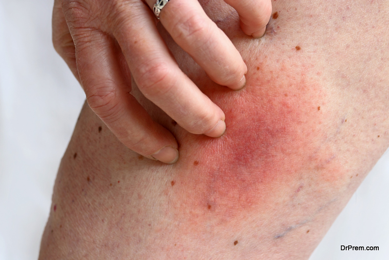
10. Physical examination
Physical examination of the patient’s body is also vital for diagnosis of squamous skin carcinoma. This test is non-invasive and requires few other laboratory tests. The diagnosis involves the examination of the skin over the whole body using a magnifying lens. The doctor looks for any unexpected growth of tissues, moles or lesions. A total-body photograph is taken to monitor changes in the skin. This test is carried out if the patient has a history of skin cancer in family, 50 or more moles on the body or symptoms of skin cancer including change in skin color, irritation or sores that do not heal. The physical examination is based on the so called ABCDE rule of examination where A stands for asymmetry in shape of moles, B for border irregularity, C for deviation in color distribution, D for diameter of the moles (larger than 6mm can be squamous skin carcinoma) and finally, E for evolution of new tissue lesions.
11. Non-invasive imaging technologies
There are different non-invasive imaging technologies that have potential to diagnose squamous skin carcinoma. These technologies include medical imaging through high frequency ultrasonography, confocal microscopy as well as optical coherence tomography. These diagnostic tests will eradicate the need of surgery to diagnose squamous cell carcinoma. The physician should strictly follow the protocols associated with these diagnostic tests. These non-invasive imaging technologies have shown significant promise but still are far ineffective when compared to skin biopsy, MRI and CT scan respectively. Moreover, these tests are not considered as standard test to diagnose squamous skin cancer.