
Bursitis: Diagnosis
Top Diagnosis
1. CT scan
A CT scan is an imaging procedure that uses a combination of X-rays and computer imaging to produce cross-sectional images of the structures inside the body. It shows detailed images of the bones, muscles, fat, and organs and is accurate for confirming or excluding bursitis. You are required to lie on a table attached to the CT scanner. The machine rotates and provides a picture of a thin slice of the affected area. This non-invasive test provides a clear understanding of the underlying problem which can further be treated efficiently.
2. Arthography
This is a specialized X-ray that views bone structures following an injection of a contrast fluid into the affected joint area. The patient lies on the screener bed as the affected part is viewed under a fluoroscope and a local anesthetic numbs the skin and tissues over the joint. A sample of joint fluid is sent to a lab to be examined under a microscope. Any abnormality observed suggests a tear, opening, or blockage in joint and possibility of a disease. An arthrogram explains what causes frequent unexplained joint pain, swelling, unusual growths, or cysts and abnormal joint movement. Care should be taken for those allergic to certain medications, dyes, local anesthesia, or iodine as the chemicals can cause infections or sometimes bleeding. The process usually takes about 30 minutes to 1 hour. Some mild pain, muscle tenderness, and swelling in joints is felt after the test. The use of ice packs, nonprescription pain relievers, and rest bring relief.
3. Crystal analysis
The need for this test usually arises after the aspiration test to look for crystals in the aspiration fluid. Crystals, if present, are examined and classified on the basis of their shape and size under polarized light. Urate and calcium pyrophosphate crystals can be easily examined through this uncomplicated lab test.
4. Aspiration test
If your doctor suspects an infection or gout in the bursa, he/she may suggest getting an aspiration test done. A sample of the bursa fluid is obtained by inserting a needle into the affected area and draining out some of the fluid through a syringe. The fluid is then evaluated under a microscope, to rule out gout or infection. Typical symptoms associated with this test are open cut wounds, redness, signs of infection such as fevers, chills, and sweats. Proper treatment can be prescribed after the fluid is tested for signs of infection. It is a semi-invasive kind of test which does not involve many complications, but patience and perseverance.
5. Ultrasound
Ultrasound or sonography, uses sound waves to produce ultrasound images of internal body parts in real time. The recorded images determine the size, shape, and consistency of soft tissues and organs. An instrument called transducer is used, which emits high frequency sounds that are inaudible to human ears. Certain precautions like not eating or drinking anything for a number of hours before the test, wearing comfortable clothing for easy screening of the affected area are required to be taken. A water-based gel is applied to the area to be screened so the transducer can easily glide over your skin making the process smoother. A technician often discusses what he observes during the test. A typical ultrasound lasts from 30 minutes to an hour. Symptoms like unusual pain in tendons, swelling and discomfort moving a particular joint are diagnosed through this test.
6. MRI
The use of radio waves and strong magnetic field which produce detailed structural images within the body is MRI. The patient in this imaging test lies on a screener bed removing all metal objects on his/her body. This non-invasive imaging test produces images of organs and structures inside the body and evidence like soft tissue swelling and fluid-filled bursa are revealed. Digital images produced from the scan can be viewed on a computer screen for further study. Problems of the bones and joints, such as arthritis, cartilage problems, bone problems, torn tendons/ligaments, infection, etc.
can be diagnosed through this extensive scan.
7. Lab tests
Laboratory tests include routine blood tests performed to confirm a particular symptom or eliminate other conditions. A blood sample is taken and studied carefully under a microscope to rule out possibility of any abnormalities and problems that can be infection of the bursa or any other cause of swelling. The test can be conducted at any pathological test center or at your doctor’s office.
8. X-ray
What the naked eye cannot see, X-rays can. X-Rays are useful in assessing a bone fracture, tumors, arthritis, or calcifications. In addition to diagnose the cause of bursitis, X-ray also reveals any other underlying problem which is causing pain. Evidence of swelling of the bursa, especially for olecranon bursitis, can also be seen through X-rays. The test can be easily conducted at pathological labs or at the doctor’s office.
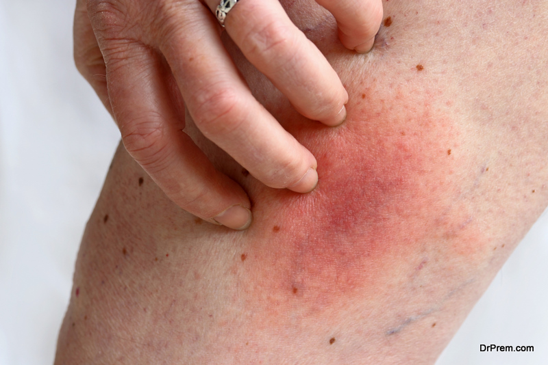
9. Physical exam
The first test doctors perform on patients with rheumatic discomfort is a physical test. Simply by pressing and observing the infected area with precision, the doctor checks for tenderness, pain, strength and range of motion in an affected area. The doctor may also assess nerve function (feeling and reflexes) and blood circulation. If the symptoms and findings indicate bursitis, you will be given treatment straightaway without any further tests.